

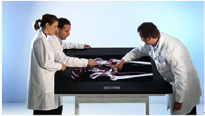
The software associated with the Visualization Table facilitates user interaction with can render 3D images of the human body. Any DICOM image can be viewed on the table. This specifically includes images generated by CAT or MRI scans as well as medical images and videos from almost any other source.
One of many online videos illustrating the Visualization Table in operation can be found here. As shown in the video, students are able to zoom in, rotate or cut into the virtual body without the use of a scalpel but in a way that, none-the-less, seems quite natural. Since the “body” is not destroyed, the same image can be used repeatedly. This not only reduces the number of bodies needed for instruction but also allows interesting or unusual conditions to be presented an unlimited number of times.

- Providing a surgeon the opportunity to become familiar with a patient’s anatomy before surgery thus allowing detailed planning of the operation.
- The touch interface allows a surgeon to interact with the virtual body with their hands, much they would during actual surgery. This enhances tactile memory and arguably increases the quality of the surgery. Furthermore, it can reduce operation and rehabilitation time by minimizing the occurrence of unexpected issues.
- The system effectively enhances collaboration between doctors.
- The system can be used to improve the quality of communications on medical issues between doctors and patients.
Closing with just a thought: given a single user and with the addition of head tracking, it would seem to be a straight forward matter to transition the system from rendered 3D imagery to active glasses-based stereoscopic 3D imagery. Doing so would provide an additional level of realism to the “off-line” medical applications described above.